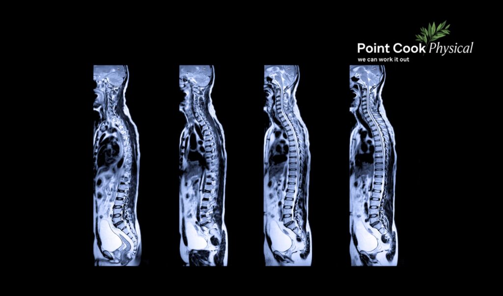
It’s common for patients to expect an MRI to provide answers for persistent shoulder pain or to question why an X-ray wasn’t the first step after a minor injury. These situations highlight a common misunderstanding: different forms of diagnostic imaging are designed to answer different questions, and choosing the right one depends on the problem being assessed.
Medical imaging isn’t one tool that works the same way for every problem. An X-ray, an ultrasound, an MRI, and a CT scan each look at the body differently, so each one answers a different question. Knowing which type of diagnostic imaging applies to your situation, and just as importantly, when no scan is needed at all, is the difference between a useful test and one that leads nowhere.
X-rays: What They Actually Show
X-rays are usually the first scan ordered, and for good reason. They use a small dose of radiation to capture dense structures like bone, which makes them the standard choice for fractures, arthritis changes, joint alignment, and bone spurs. A specialised version, the DEXA scan, measures bone density and is used to assess osteoporosis risk.
What an X-ray doesn’t show well is soft tissue. Muscles, tendons, ligaments, and cartilage barely register on this type of imaging. A normal X-ray after a sprained ankle or a sore shoulder doesn’t mean nothing is wrong. It often means the problem sits in tissue this particular scan was never designed to capture.
A practical example: someone who falls and suspects a wrist fracture will typically get an X-ray first. Someone with persistent shoulder pain but a clean X-ray usually needs a different type of scan to find what’s actually going on.
Ultrasound: A Close Look at Soft Tissue in Real Time
Ultrasound uses sound waves rather than radiation, which makes it well suited to tendons, bursae, and muscles close to the skin’s surface. Because the images appear in real time, a sonographer can also watch how a tendon or joint behaves during movement, something a still image can’t offer.
This is why ultrasound is a common first step for rotator cuff tendinopathy, tennis elbow, bursitis, and Achilles tendon problems. It’s also used to guide injections, such as cortisone or PRP, directly to the affected area.
Ultrasound has limits too. It struggles to see deep into joints, through bone, or into complex internal structures. A shoulder with suspected deep joint damage may need an MRI instead. For straightforward soft tissue injuries close to the surface, ultrasound is often the most practical and cost-effective imaging option.
MRI: The Detailed Option for Soft Tissue and Nerves
MRI uses magnetic fields rather than radiation, producing detailed images of tendons, ligaments, cartilage, nerves, muscles, and bone marrow all in one scan. In an MRI vs ultrasound comparison, the difference comes down to depth and detail. MRI reaches structures ultrasound can’t, including deep inside a joint or within the bone itself.
This makes MRI the preferred scan for rotator cuff tears, meniscus tears in the knee, ACL injuries, and stress reactions that haven’t yet appeared on an X-ray. It also comes into play when someone has persistent, unexplained joint pain and earlier imaging hasn’t provided an answer.
The trade-offs
MRI costs more than an X-ray or ultrasound, appointment wait times tend to be longer, and the scan itself takes longer to complete. For these reasons, MRI is usually kept for cases where the diagnosis genuinely needs that level of detail, rather than being the first scan ordered for every injury.
CT Scans: Detailed Bone Imaging for Complex Cases
CT scans combine multiple X-ray images into a detailed, three-dimensional view of bone. They earn their place where a standard X-ray falls short, particularly with complex fractures, fractures involving a joint surface, or injuries where a surgeon needs precise detail before planning an operation. CT is also useful for tracking how a fracture is healing over time.
What CT doesn’t do particularly well is show soft tissue detail, which is where MRI takes over. In practice, the two scans tend to be complementary rather than competing options. A complex wrist or pelvic fracture might call for a CT scan, while a ligament tear in the same general area points toward MRI instead.
Bone Scans: Finding What Other Scans Miss
A nuclear medicine bone scan works differently again. A small amount of radioactive tracer is injected, which travels to areas of increased bone activity. This makes bone scans useful for detecting stress fractures, infection, or other bone changes that haven’t shown up on X-ray, particularly when pain persists despite normal imaging.
A related test, SPECT-CT, combines this bone scan information with a CT scan’s anatomical detail to pinpoint the exact source of pain. It can help with unclear spinal or foot pain after other scans haven’t given a clear answer. Bone scans aren’t requested often in general musculoskeletal care, but they fill a specific gap: identifying bone-related problems that other imaging has missed.
Why Scanning Often Isn’t the First Step for Back Pain
Back pain is the condition people most expect a scan for, and it’s also the condition where a scan is least likely to change the outcome. Clinical guidelines consistently point out that imaging findings such as disc bulges or mild degeneration show up just as often in people without any back pain at all. A scan may reveal changes or findings that are not necessarily the cause of your symptoms.
This is part of why a thorough physiotherapy assessment typically comes before any imaging request. A detailed history and physical examination tells a practitioner more about what’s driving your pain than a picture of your spine, and acting on an incidental finding from a scan can send treatment down the wrong path entirely.
Imaging becomes appropriate for back pain when conservative treatment hasn’t helped after a reasonable period, when symptoms are getting worse rather than better, when there are red flag signs such as unexplained weight loss, fever, or nerve symptoms, or when the clinical picture genuinely doesn’t add up. Outside those situations, a scan is more likely to create confusion than clarity, sending you chasing a finding that may have nothing to do with how you feel.
When You Should Get a Scan, and When You Shouldn’t
A scan is generally worth pursuing when one or more of these apply: conservative treatment has been given a fair trial and hasn’t worked, symptoms are progressively worsening rather than settling, your practitioner is no longer confident in the working diagnosis, or there’s suspicion of something more serious, like infection, fracture, or a tumour.
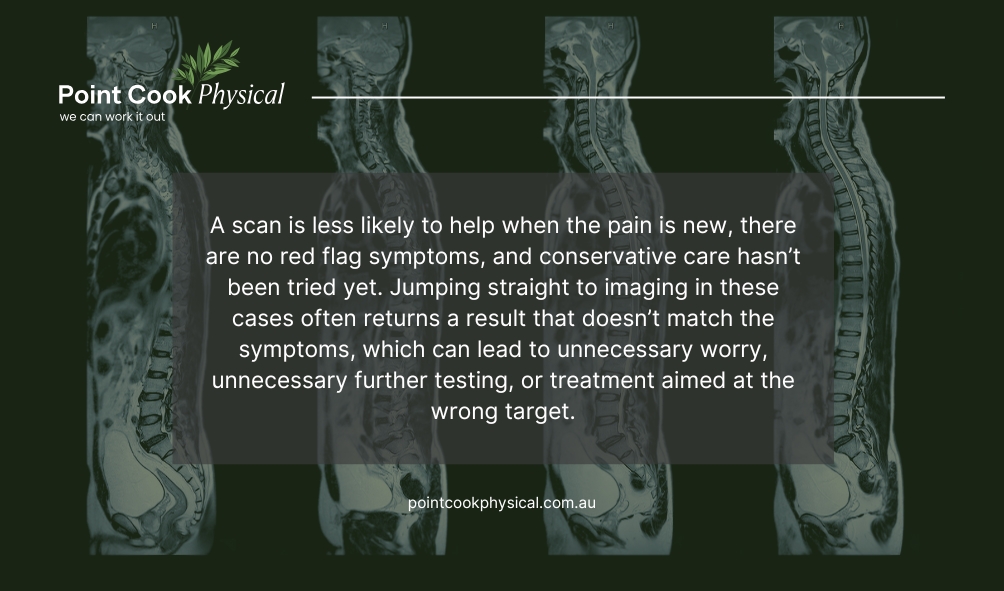
Diagnostic imaging is less likely to help when the pain is new, there are no red flag symptoms, and conservative care hasn’t been tried yet. Jumping straight to imaging in these cases often returns a result that doesn’t match the symptoms, which can lead to unnecessary worry, unnecessary further testing, or treatment aimed at the wrong target.
This is a decision best made with your treating practitioner rather than worked out alone, since the right type of diagnostic imaging, or whether any imaging is needed at all, depends on your specific history, examination findings, and how your symptoms are tracking over time.
A quick rule of thumb: what scans serve what purpose
- Bone problem suspected → start with an X-ray.
- Tendon or bursa problem → ultrasound, often first.
- Ligament, cartilage, muscle, nerve, or unclear pain source → MRI.
- Complex fracture or surgical planning → CT.
- Hidden bone injury or unexplained bone pain → bone scan.
Medical Imaging For Injuries: Your Questions Answered
| Question | Answer |
|---|---|
| What’s the difference between an X-ray and an MRI? | An X-ray uses radiation to capture bone detail and is the usual first scan for fractures, arthritis, and alignment problems. An MRI uses magnetic fields to show soft tissue such as tendons, ligaments, cartilage, and nerves in far greater detail, making it the better option when bone imaging alone hasn’t explained the pain. |
| Why don’t I need a scan for my back pain? | Most back pain improves with conservative care, and scans often show findings, like disc bulges, that are common even in people without pain. A thorough physiotherapy assessment usually identifies the cause more reliably than imaging, with scans reserved for cases that aren’t improving or that show red flag symptoms. |
| When should I get a scan for an injury? | A scan is generally worthwhile when conservative treatment hasn’t helped after a reasonable period, symptoms are worsening, the diagnosis remains unclear, or there are signs of something more serious. Outside these situations, a clinical assessment is usually the more useful first step. |
| Is ultrasound or MRI better for a soft tissue injury? | Ultrasound works well for tendons, bursae, and muscles close to the skin and allows real-time movement assessment, often at a lower cost. MRI provides more detail for deeper structures, including ligaments, cartilage, and nerves, and is typically used when ultrasound findings are inconclusive. |
| Do I need a referral to get a scan? | Most imaging, including X-rays, ultrasounds, MRIs, and CT scans, requires a referral from a GP, physiotherapist, or other treating practitioner. This ensures the right scan is chosen for your specific presentation rather than requesting imaging that may not answer the clinical question. |

Final Thoughts
There’s no single best scan, only the right scan for what’s actually going on. X-rays handle bone, ultrasound and MRI handle soft tissue at different depths, CT handles complex bone detail, and bone scans pick up what the others miss. Back pain sits slightly outside this picture, since a careful assessment usually tells your practitioner more than a scan will.
If you’re unsure whether your injury or pain needs imaging, that question is worth raising with your physiotherapist or osteopath before booking a scan yourself.
Book an assessment at Point Cook Physical and we’ll help work out whether a scan is actually going to add anything to your treatment, or whether it’s a step you can skip altogether.